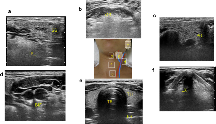
Fig. 1.
Basic image of the neck (systematic cervical ultrasonography). SG submandibular gland, PL: pharyngeal lateral wall, OB: oral base, PG: parotid gland, BIF: carotid bifurcation, TR: trachea, TH: thyroid gland, ES: esophagus, LX: larynx. The probe should be continuously scanned to ensure that it passes through these sites, and the left and right necks should always be observed. Observe in a manner that does not overlook changes in lymph nodes, blood vessels, and muscles along the way. a Submandibular region (submandibular gland, facial arteriovenous, oropharyngeal sidewall). b Submental region (oral floor muscles, base of tongue). c Parotid region (parotid gland, mandible, masseter muscle). d Carotid bifurcation (external carotid artery, internal carotid artery, internal jugular vein). e Anterior neck (thyroid, common carotid artery, internal jugular vein, cervical esophagus). f Larynx

