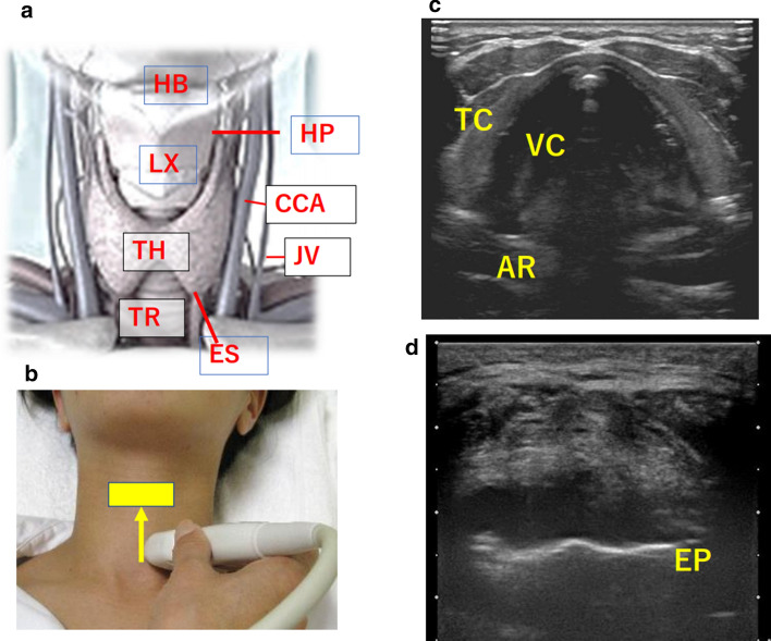
Fig. 2.
POCUS of the anterior neck HB hyoid bone, LX larynx, TH thyroid gland, TR trachea, HP hypopharynx, ES esophagus, CCA common carotid artery, JV internal jugular vein, VC vocal cord, TC thyroid cartilage, AR arytenoid cartilage, EP epiglottis cartilage. a Anterior neck anatomy. b Anterior neck probe operation. c Transverse view of the anterior neck: Ultrasound image of the larynx (vocal cord level). d Transverse view of the anterior neck: Ultrasound image of the larynx (supraglottic level)

