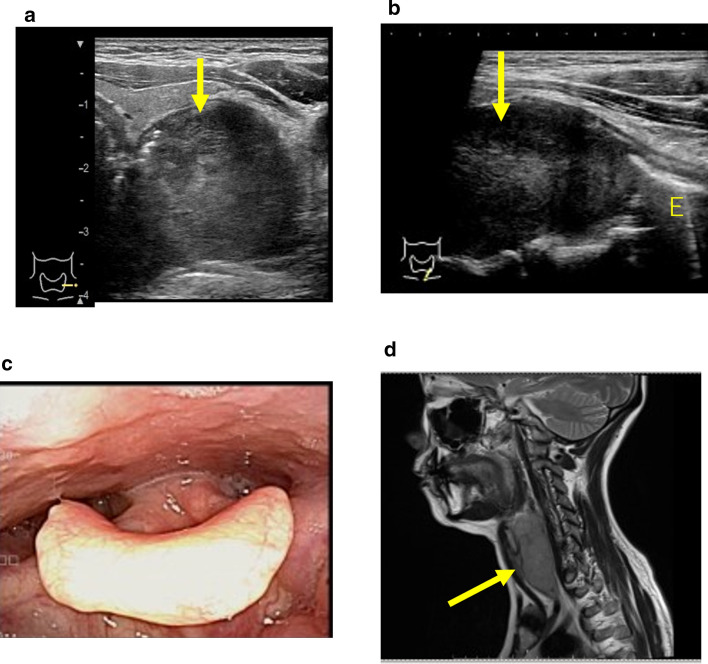
Fig. 4.
Advanced cancer of the cervical esophagus. a Transverse view of the lower part of the anterior neck: A tumor (↑) was found on the dorsal side of the left lobe of the thyroid gland. b Sagittal image of the lower part of the anterior neck: The tumor (↑) led to the cervical esophagus (E). c Laryngeal fiberscope findings: No obvious neoplastic lesions can be pointed out. d Cervical MRI findings (sagittal section): A tumor (↑) was found from the posterior hypopharyngeal ring to the cervical esophagus

