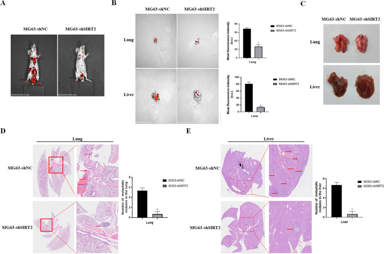
Fig. 6. SIRT2 knockdown inhibited the metastatic ability of osteosarcoma cells in vivo.
A Small animal in vivo imager was used to detect the metastasis of osteosarcoma cells in the SIRT2 knockdown group and the control group. B Fluorescence images of the lung and liver in the SIRT2 knockdown group and control group. A representative fluorescence image of at least three tissue samples is shown. The right panel is the summarized data of immunofluorescence density. C Photographs of the lung and liver of the two groups of nude mice. D, E H&E staining of paraffin-embedded lung and liver sections. The right panel is a magnification of the rectangle in the left panel, with arrows showing metastatic nodules. Number of metastatic nodules in the lung or liver is summarized in a histogram format. (*P < 0.05, **P < 0.01).

