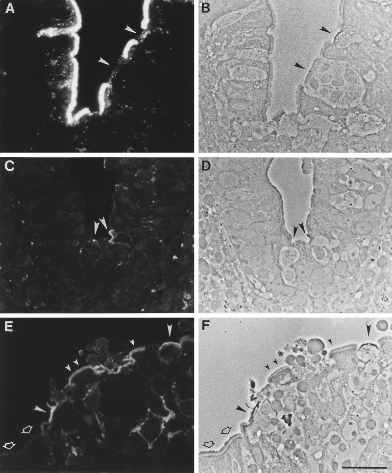
FIG. 1.
Lectin and antibody binding patterns in normal human Peyer’s patch. Sections were labeled with lectin or antibody and viewed by fluorescence (A, C, and E) and phase-contrast (B, D, and F) microscopy. (A to D) A dome with FAE is on the right, and a villus is on the left. (A and B) UEA I lectin, which selectively labels BALB/c mouse PP M cells, has abundant binding sites expressed uniformly on human enterocytes. Conversely, little or no label is seen on M cells in the FAE (arrowheads). (C and D) Monoclonal antibody specific for sialyl Lewis A antigen shows selective binding to apical membranes of M cells (arrowheads) as well as weaker staining of internal and basolateral membranes. (E and F) On the FAE, anti-sialyl Lewis A strongly labels most M cells (large filled arrowheads). A small (<20%) population of FAE enterocytes (small filled arrowheads) are also labeled. However, most enterocytes are negative (open arrowheads). Bar, 20 μm.

