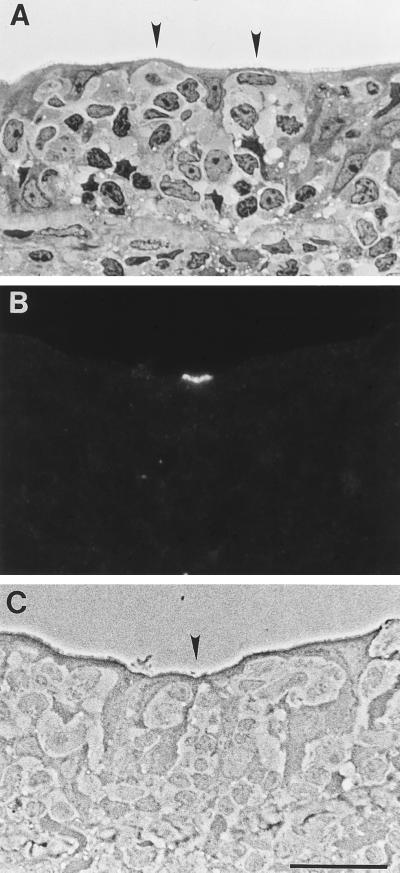
FIG. 3.
Anti-sialyl Lewis A binding pattern in normal human cecum. Sections were stained with toluidine blue and viewed by brightfield microscopy (A) or labeled with anti-sialyl Lewis A and viewed by fluorescence (B) and phase-contrast (C) microscopy. In cecal specimens which contained lymphoid follicles, identifiable M cells were more abundant in the FAE than in Peyer’s patch. (A) M cells (arrowheads) were clustered in certain regions of the FAE. (B and C) Anti-sialyl Lewis A labeled the apical membranes of a small subpopulation of M cells (arrowhead). No other cell types were recognized by this probe. Bar, 20 μm.

