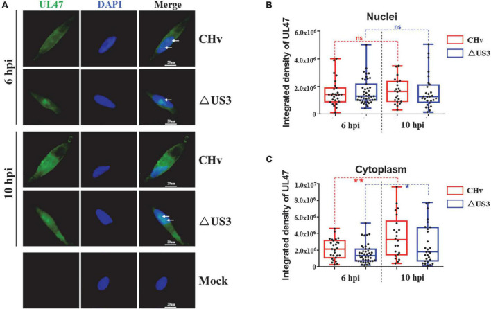
FIGURE 7.
The cytoplasmic translocation of UL47 was delayed in ΔUS3 infection. DEF cells were infected with two MOI of CHv and ΔUS3. Infected cells were fixed at 6 and 10 hpi, and UL47 protein and the nuclei were labeled. (A) Lack of US3 protein impaired UL47 diffusion in the nuclei at the late stage of infection. White arrows indicate UL47 aggregates. (B) The nuclear UL47 density in CHv and ΔUS3 infection. (C) The cytoplasmic UL47 density in CHv and ΔUS3 infection. The ImageJ software was used to analyze the integrated density of UL47 in the whole cells and the nuclei. The data were presented as Min. to Max. with the mean and all points using GraphPad Prism 6 software. *P < 0.05; **P < 0.01. ns, no significant difference.

