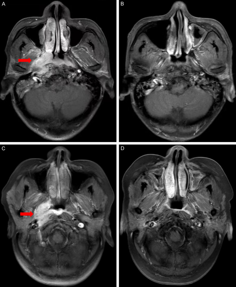
Figure 2.

MRI of nasopharyngeal lesions in experimental group and control group before and after treatment. Representative MRI images before and after treatment for experimental and control group. (A) Baseline MRI of nasopharyngeal lesions in a 56-year-old man with nasopharyngeal carcinoma in the experimental group, (B) Reexamination of MRI after treatment showed complete response (CR). (C) Baseline MRI of nasopharyngeal lesions in a 53-year-old woman with nasopharyngeal carcinoma in the control group, (D) Reexamination of MRI after treatment showed complete response (CR).
