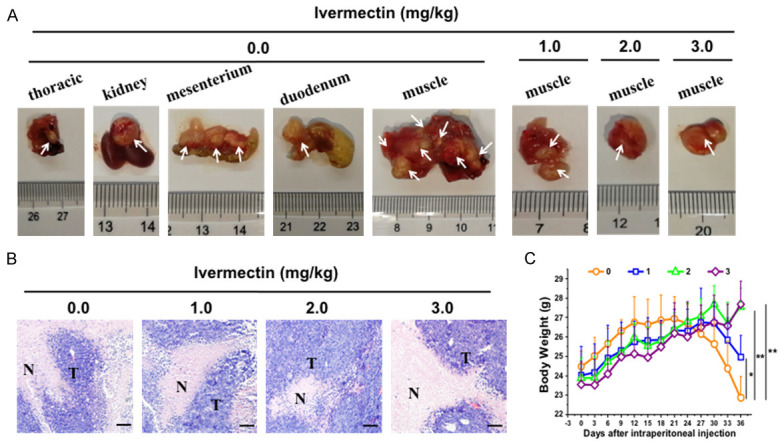
Figure 3.

IVM inhibited the metastasis of HCT-8 cells-derived tumor in vivo. The NOD/SCID mice were injected through tail vein with 2 × 10⁶ HCT-8 cells. The mice were then treated with different doses of IVM (0, 1, 2, or 3 mg/kg, i.p.) daily for 37 days. A. The mouse tissues that contained tumor nodules. White arrows indicated the metastatic tumor nodules. B. The histochemicl examination of the tumor mass found in muscle tissue with hematoxylin and eosin staining. Scale bars = 150 μm. N, non-tumor tissues; T, tumor tissues. C. The comparison of the body weight of the HCT-8 cells-inoculated mice after IVM treatment. The numbers 0, 1, 2, and 3 in the figure keys represented the IVM doses 0, 1, 2, and 3 mg/kg, respectively. Data were presented as the mean ± SD (n = 6). *P < 0.05, **P < 0.01, compared with the vehicle controls.
