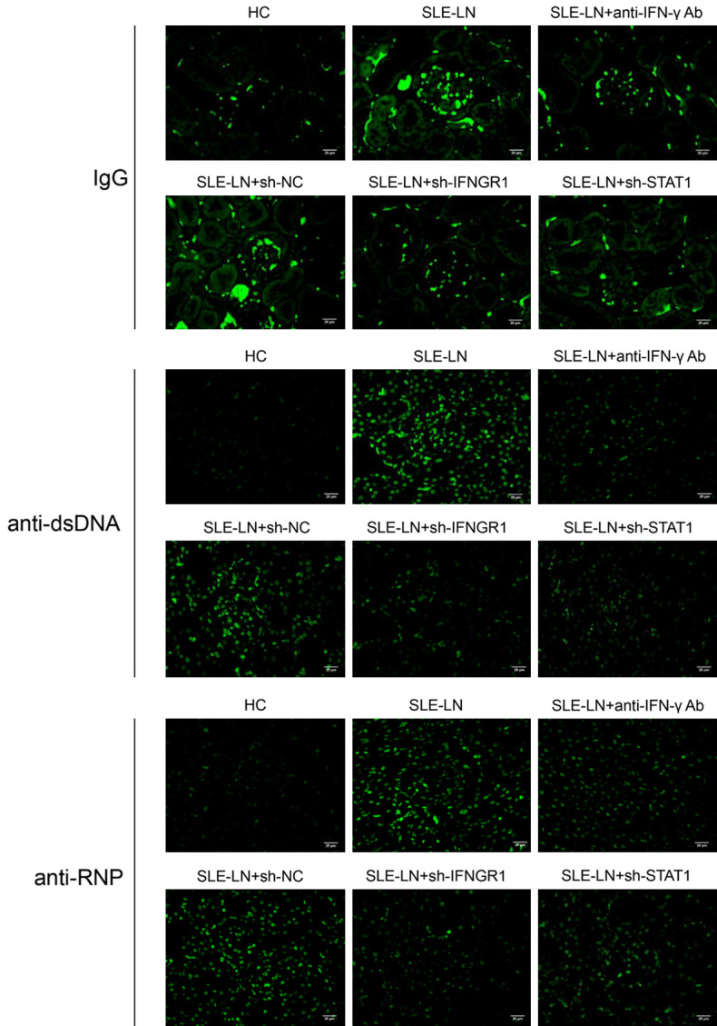
Figure 7.

Immunofluorescent analysis of IgG, anti-dsDNA and anti-RNP levels in kidney sections from mouse SLE models. Sections (mouse kidney tissue) from the indicated groups were stained for IgG, anti-dsDNA and anti-RNP. SLE: systemic lupus erythematosus; HC: healthy controls; LN: lupus nephritis; NC: negative controls. (Magnification: 400X, scale bar: 20 μm).
