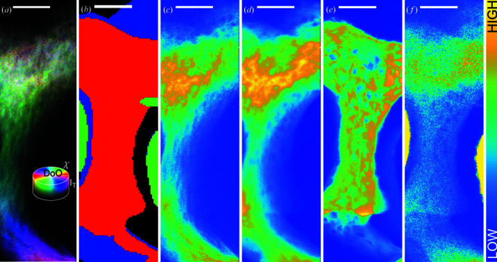
Figure 5.
Analysis of the test sample consisting of a tensile load screw and surrounding bone. (a) Orientational distribution H(ue) S(aturation) V(alue) map of the full sample, for every pixel the process like that described in Section 2.2.2 has been carried out if sufficient HAP (002) signal was present. The hue of a point reveals the crystallite orientation, the saturation reveals the projected degree of orientation and the value reveals the total projected (002) intensity in the point. (b)–(f) XRF and XRD analysis. (b) Segmentation sketch based on XRF signals. The Ca signal in red shows areas where bone is close to the sample surface, the Ni signal in green shows the location of the implant tension spring, and the Sr signal in blue shows areas where there is bone further from the sample surface. (c) HAP (002) integrated intensity. (d) HAP multiplet integrated intensity. (e) XRF Ca signal. (f) XRF Sr signal. Scale bars are 50 µm. The outermost 3 µm in (a)–(f) have been removed on both the left and the right, due to edge effects; thus (a), (c) and (d) contain 104 × 350 diffractograms.

