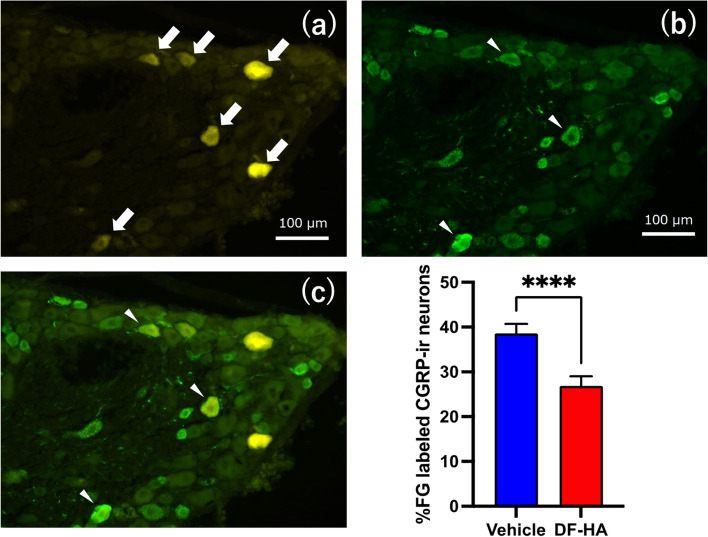
Fig. 2.
Fluorescence photomicrographs of the DRG after administration of DF-HA or vehicle for 4 weeks a FG-labeled DRG neurons, b CGRP-immunoreactive (ir) DRG neurons, and c overlaid picture a on b. All photomicrographs are from the same section. White arrows in the photomicrograph of a indicate FG-labeled DRG neurons, and white arrowheads in the photomicrograph of b and c indicate FG-labeled CGRP-ir DRG neurons. The proportions of FG-labeled CGRP-ir neurons in the DF-HA groups are significantly higher than those in vehicle group (n = 4 rats per group)

