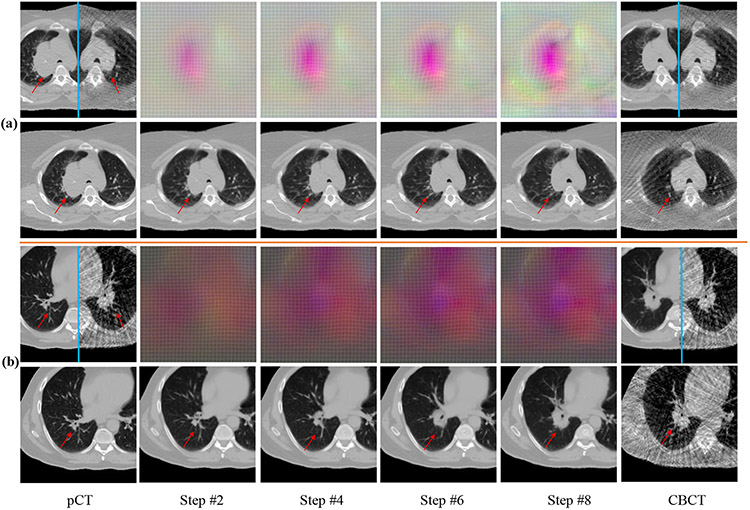
Fig. 5.
Progressive DVFs with warped pCTs (rows 2 , 4) for a shrinking tumor (row 1), and out of plane rotation (row 3). Mirror flipped view of pCT and the CBCT before and after alignment are shown. DVF colors indicate displacements in x (0mm to 10.13mm) (black to red),y (0mm to 7.76mm) (black to green), and z (0mm to 13.50mm) (black to blue) directions. Red arrow identifies the tumor.

