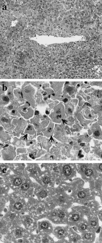
FIG. 5.
Histology of hepatic injury in mice injected with d-GalN and LPS. Mice were injected i.p. with the mixture of d-GalN and LPS, and livers were removed from live mice 12 h after the injection. Liver sections from mice injected with d-GalN and LPS (a and b) or saline alone (c) were stained with hematoxylin and eosin. Note hepatic injuries around a blood vessel (a) and fragmented nuclei of hepatocytes (arrows in panel b). Magnifications: a, ×200; b and c, ×1000.

