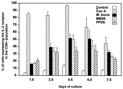
FIG. 3.
Kinetics of activation of CD8+ T cells. PBMC from M. bovis-infected animals (01, 84, and 97) were stimulated in vitro with live M. bovis (106 CFU/ml), MBSE (4 μg/ml), or PPDb (4 μg/ml) for different periods (1.5, 3.5, 4.5, 6.5, and 7.5 days). Control (no-antigen) and concanavalin A (Con A)-stimulated cultures were also included. Activation of CD8+ T cells within the short-term cultures was determined by staining for CD8 and IL-2R at each of the time points and analyzed by flow cytometry. Results are representative of two experiments and are expressed as the mean value from the three animals ± standard error.

