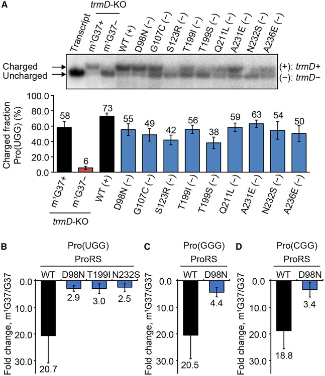
Figure 3. Prolyl-aminoacylation of proS suppressors is less dependent on m1G37.
(A) Acid-urea gel of cellular prolyl-aminoacylation status of each reconstructed suppressor. An uncharged and un-modified Pro(UGG) is in lane 1, a pair of control tRNA samples of MG1655-trmD-KO grown in m1G37+ and m1G37− conditions in lanes 2 and 3, and the WT MG1655 sample is in lane 4. Data are mean ± SD (n = 3).
(B–D) Kinetics of prolyl-aminoacylation of (B) Pro(UGG), (C) Pro(GGG), and (D) Pro(CGG), showing loss of kcat/Km of the WT enzyme upon removal of m1G37 relative to proS mutants. Data are mean ± SD (n = 3).

