
Fig. 2.
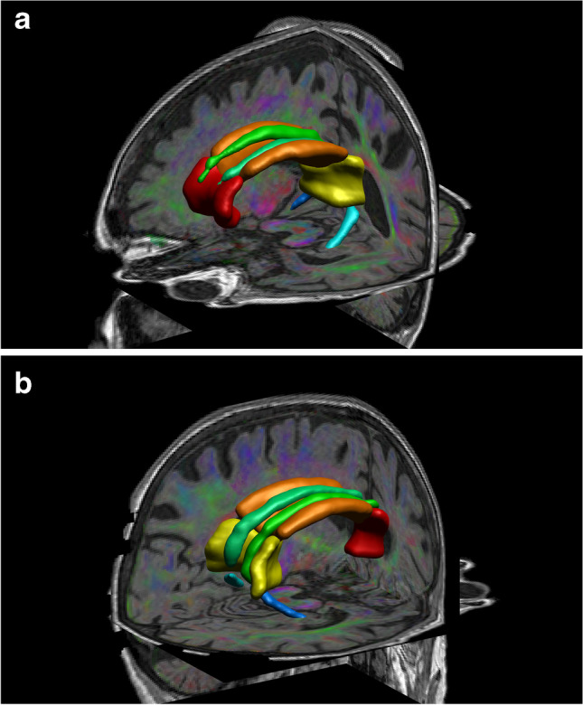
a 3D left anterior–posterior view; b 3D right posterior-anterior view of one study subject’s regions of interest projected over the fused colour coded fractional anisotropy and T1 weighted images. Red = genu of corpus callosum, orange = body of corpus callosum, yellow = splenium of corpus callosum, emerald green = right cingulate gyrus, jade green = left cingulate gyrus, dark blue = right parahippocampal cingulum, light blue = left parahippocampal cingulum