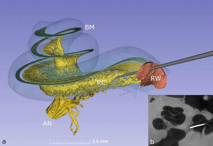
Figure 1.
(a) Synchrotron phase contrast imaging (SR-PCI) with 3D orthographic rendering of an intact left human inner ear. The bony wall of the cochlea was made semi-transparent to permit visualization of the basilar membrane (BM), Rosenthal’s canal (RC) and auditory nerve (AN). The auditory nerve contains approx. 30,000 fibres and their cell bodies are located in a 14.5 mm long spiral bony canal called RC. From there, peripheral neurites spread out to innervate approx. 15,000 hair cells placed on the BM. A probe is shown penetrating the round window (RW) membrane to access the underlying RC. (b) Microradiograph taken following placement of a radio-opaque marker at the presumed site of RC on anatomical dissection; the image confirms precise targeting of RC during dissection.

