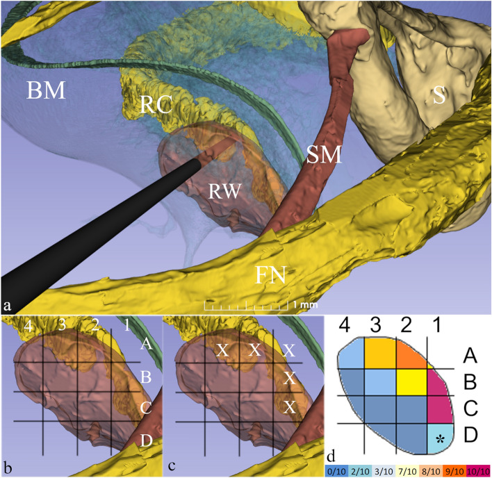
Figure 2.
(a) The surgical approach to left Rosenthal’s Canal (RC) passing through the mastoid bone behind the ear. Note the structures of surgical interest: the facial nerve (FN), the stapes (S) and stapedius muscle (SM) and round window membrane (RW). A surgical trephine is seen penetrating the RW to reach RC located just deep to it. (b) A 4 × 4 dynamic grid was applied to 10 RW membranes and was adjusted to membrane size for each specimen. (c) X denotes those grid units in closest anatomical proximity to RC in which a given penetration had at least an 80% chance of reaching RC (see Supplementary Table 2). (d) Using this data, a heat map was created for optimal targeting of RC behind the RW membrane. Highest probability of targeting the RC was through the superior mid-region of the RW membrane. At A2 and A3, the chance was 90% and 80% respectively. Higher values were noted in C1 and B1 (100%), but this region risks injury to the vestibulo-cochlear artery. *D1 was covered by stapedius muscle and was thus not evaluable. Colour bar at the bottom displays the actual frequencies of successfully targeting the RC.

