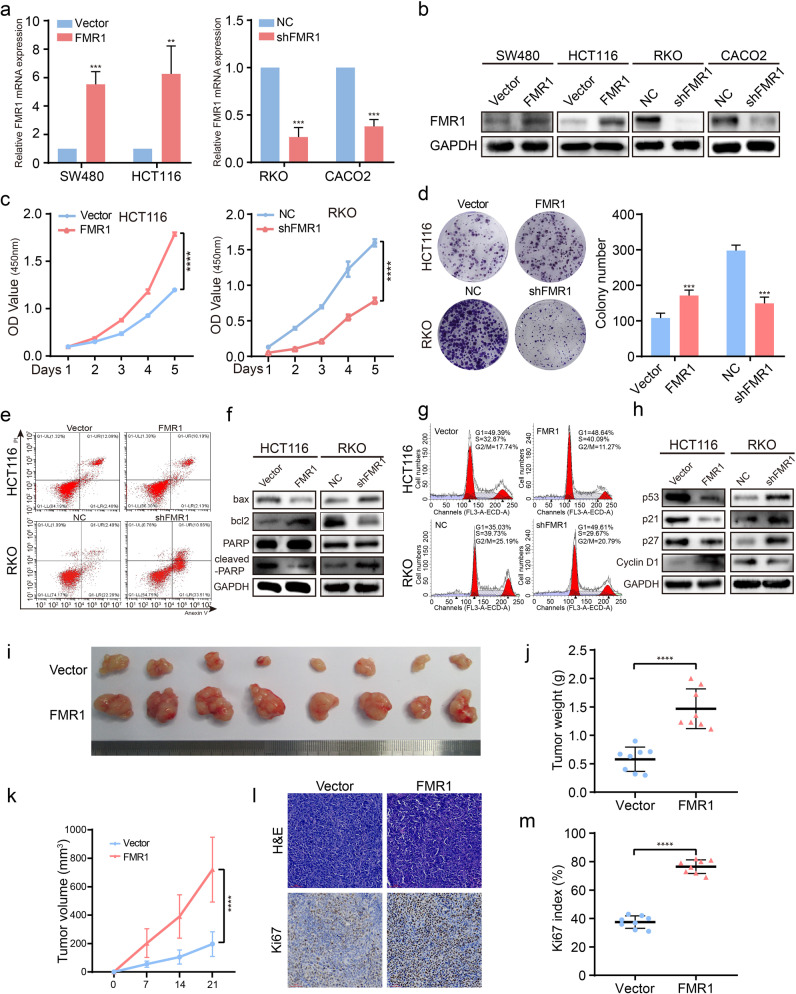
Fig. 2. FMR1 regulates proliferation, apoptosis, and cell cycle of CRC cells in vitro and in vivo.
a RT-qPCR was used to validate the expression of FMR1 in SW480 and HCT116 CRC cells with FMR1 stable overexpression and knockdown. b Western blot was used to validate the expression of FMR1 in CRC cells with FMR1 stable overexpression and knockdown. c CRC cell proliferation was analyzed by CCK8 assays. d Representative results of colony formation; the numbers of colonies containing >50 cells were scored. The number of colonies counted was of an entire well and the error bars represent mean ± SD from three independent experiments. e Apoptosis assay by flow cytometry. Annexin-positive/PI-negative (right lower quadrant) cells were analyzed for apoptosis rate. f Western blot was used to test the molecular markers of apoptosis. g Flow-cytometry analyses of the cell cycle of the indicated CRC cells. h Western blot was used to test the molecular markers of cell cycle. i The xenograft models were generated after injecting HCT116/Vector and HCT116/FMR1 cells in nude mice (n = 8/group). j The tumor weight was measured after the nude mice were euthanized. Error bars represent the means ± SD. k The tumor volumes were measured on the indicated days. The data points represent the mean tumor volumes ± SD. l The sections of tumor were subjected to H&E staining or IHC staining using an antibody against Ki-67. m Ki67 index was calculated. The data points represent the mean tumor volumes ± SD. **P < 0.01, ***P < 0.001, ****P < 0.0001.

