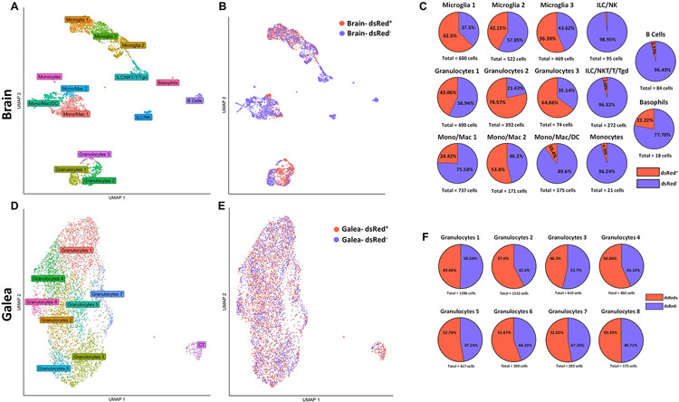
Figure 3. scRNA-seq identifies diverse transcriptional clusters of phagocytic vs. non-phagocytic CD45+ cells in the brain and galea during S. aureus craniotomy.
C57BL/6J mice (n=25) were sacrificed at day 3 following craniotomy infection with a S. aureus-dsRed strain, whereupon viable phagocytic (dsRed+; n= 1,757 in the brain and n= 2,878 in the galea) vs. non-phagocytic (dsRed−; n=2,563 in the brain and n= 2,510 in the galea) CD45+ cells were purified by FACS for scRNA-seq. Uniform manifold approximation and projection (UMAP) plots depicting transcriptional clusters in the (A) brain and (D) galea that were further separated into phagocytic and non-phagocytic cells in the (B) brain and (E) galea. The percentages of dsRed+ vs. dsRed− cells in the (C) brain and (F) galea are presented.

