
Fig. 3.
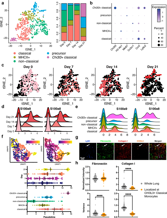
Tumor-associated monocytes in late-stage disease support ECM remodeling. a Five monocyte phenotypes were identified in the lung and their populations were time dependent. b Classical and non-classical (alternatively activated) monocytes were identified through Ccr2 and Cx3cr1 expression. c Monocyte heterogeneity decreased as time progressed. Pink dots identify cells at the designated time point. d Time-dependent responses of S100a8 and S100a9 in the monocytes. e Responses of S100a8 and S100a9 in each of the monocyte subsets. f Comparison of the real-time monocyte progression to pseudotime analysis. The star indicates the pseudotime origin as predicted by the Monocle3 algorithm. g Immunofluorescence staining of monocyte/ECM-related proteins in lung sections at day 21, blue = Ly6C, green = fibronectin, red = collagen I, orange = CHI3L3. Arrows point to CHI3L3 + monocytes. h Quantification of fibronectin and collagen I intensity and variance in the lung and in the area immediately surrounding CHI3L3 + monocytes (refer to Fig. S7 for determination of localized area), ****p < 0.0001