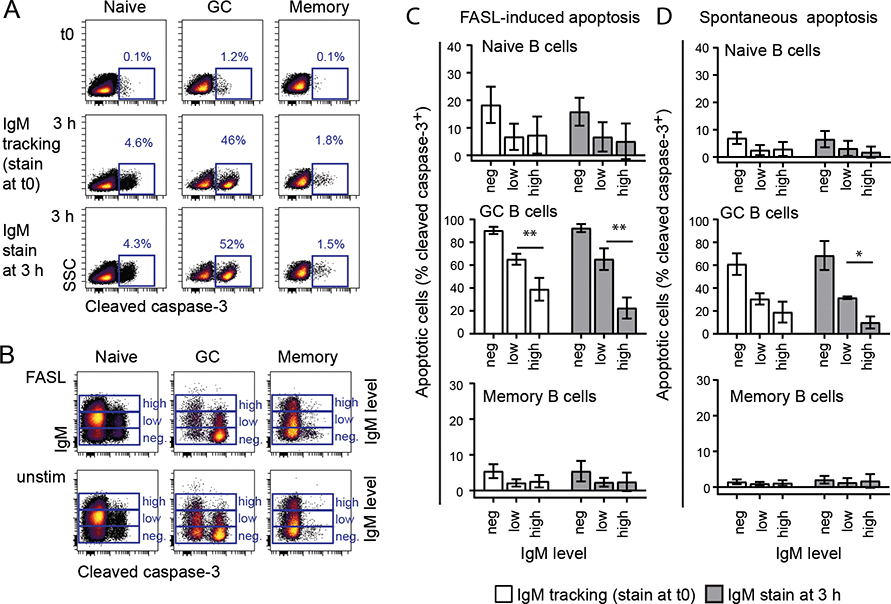
Figure 2. High BCR surface expression is associated with low spontaneous and FASL-induced apoptosis.
Single cell suspensions from human tonsils were thawed and cultured for 3 hours with or without FASL. Dead cells were excluded by a live/dead marker and apoptosis was measured by anti-cleaved caspase-3 antibody using flow cytometry. Cells were either stained with anti-IgM F(ab’)2 after fix at the end of the 3-hour culture or prior to culturing; 5 min stain and wash before culturing (“IgM tracking”). Gating of B-cell subsets were performed as in Figure 1. A. IgM-tracking did not affect spontaneous apoptosis in unstimulated naive, memory or GC B cells. B-D. B cells were divided in three populations based on IgM level and percentage of cleaved caspase-3+ apoptotic cells were gated as in A. Mean ± SD, n = 3. *indicates significance in a t-test between high and low IgM expression. *p < 0.05, **p < 0.01.

