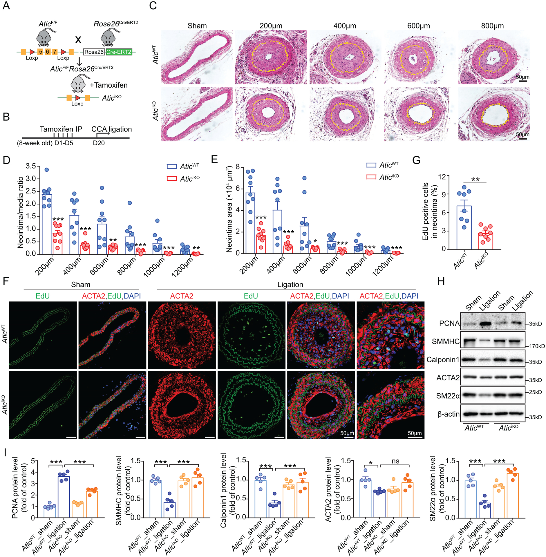
Figure 6. Global Atic deficiency attenuates neointima formation in a model of vascular injury.

(A) Strategy for generating AticiKO mice by crossing AticF/F mice with Rosa26Cre/ERT2 mice and tamoxifen treatment of the consequent mice. (B) Schematic timeline of tamoxifen treatment and carotid artery ligation model. (C) Representative HE-stained cross-sections of carotid arteries from the mice with or without left common carotid artery ligation for 28 days. Sections are 200 to 800 μm from the site of ligation. Yellow lines indicate the internal elastic lamina. (D-E) Quantification of the arterial neointima area and the ratio of neointima area to medial area of injured carotid arteries. (n = 9). (F-G) Representative EdU-positive stained cells in the neointima of carotid arteries from AticWT and AticiKO mice and their quantification (n = 8). (H-I) Representative Western blot (H) and quantification (I) of the indicated proteins normalized to β-actin in the sham and ligation-injured carotid arteries of AticWT and AticiKO mice 7 days post ligation injury (n = 5). Data are represented as mean ± SEM, *P < 0.05, **P < 0.01 and ***P < 0.001 for indicated comparisons.
