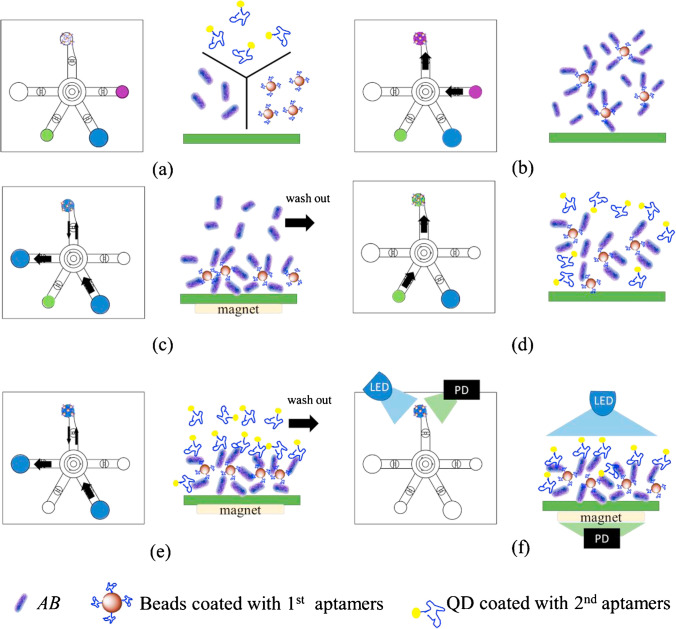
Fig. 9.
Dual aptamer assay for the detection of AB. (a) Bacterial samples and reagents were loaded into the corresponding chambers. (b) Magnetic beads and bacteria were pumped into the reaction chamber by a micropump and mixed by a micromixer. (c) Unbound materials were washed away with wash buffer while applying an external electromagnetic field. (d) Bead-bacteria complexes and QD were mixed by a micromixer. (e) Excessive QD was washed away with wash buffer while applying an external electromagnetic field. (f) Fluorescent signals were excited by LED and detected by photodiodes (PD). Reproduced with permission from [122], Copyright 2020 Elsevier

