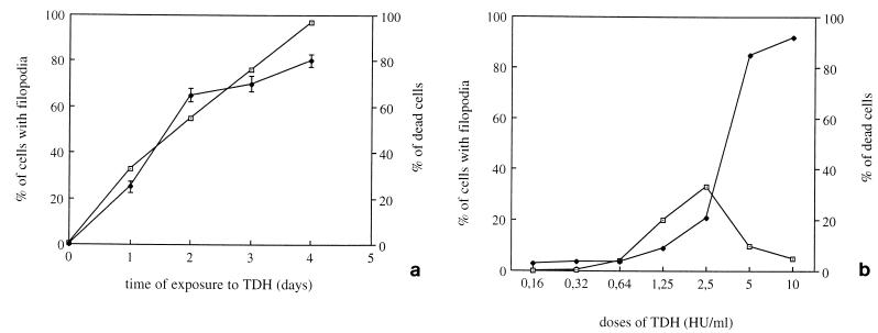
FIG. 5.
TDH-induced effects in IEC-6 cells are dose and time dependent. (a) Percentages of cells with filopodia and of dead cells after various times of exposure to 2.5 HU of TDH per ml; (b) dose dependence of TDH-induced cellular effects (filopodium formation and cell death), detectable after 18 h of exposure to the toxin. The results reported as percentages (± standard deviations) of cells with filopodia (⊡) or of dead cells (⧫) (as detected by trypan blue) with respect to the total number of cells counted, are from three different experiments in each of which at least 500 cells (randomly chosen) were counted.

