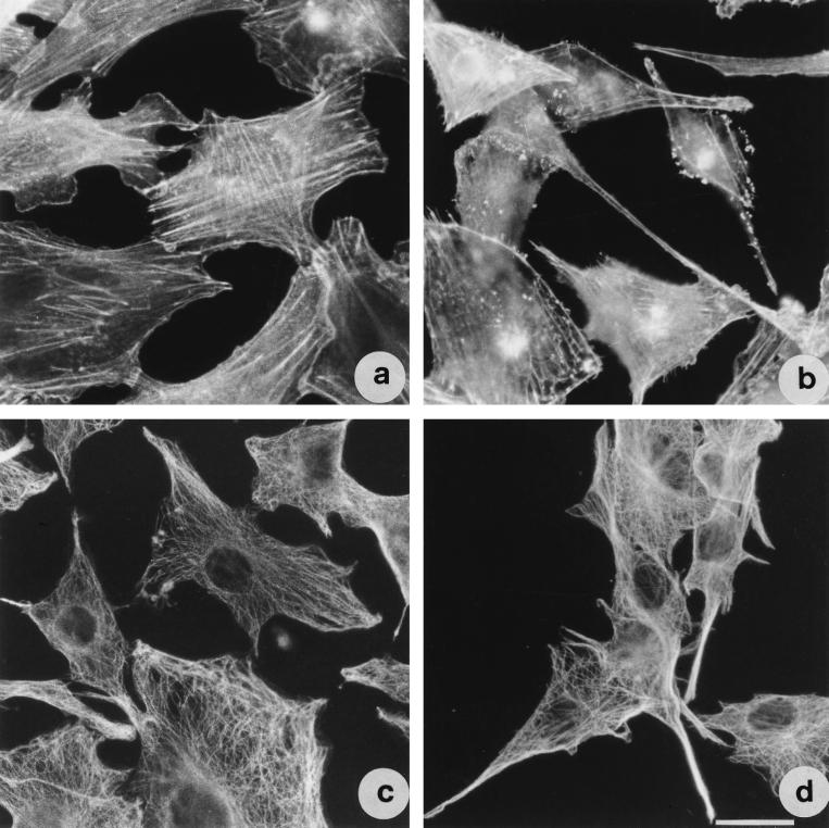
FIG. 7.
TDH provokes changes in actin and tubulin cytoskeletal networks in IEC-6 cells. Shown are fluorescence micrographs of cells stained for detection of F-actin (a and b) and tubulin (c and d). (a and c) Control cells; (b and d) cells exposed to 2.5 HU of TDH per ml for 18 h. Both cytoskeletal elements are present in the filopodia. Bar represents 10 μm.

