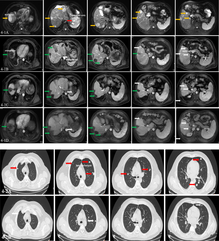
Figure 4.
Imaging of clinical events. A man aged 45 years was diagnosed as having moderately differentiated hepatocellular carcinoma at Barcelona Clinic of Liver Cancer (BCLC) C stage, who received the combination therapy. (4-1A) The T2 phase of MRI images showing multiple lesions in the liver and abdominal cavity (yellow arrow: liver lesions; red arrow: abdominal cavity metastasis). (4-1B) The arterial phase of MRI images showing multiple ablated zone (green arrow), subtotal ablation of the lesions, and unablated lesion in the liver (white arrow) at baseline. (4-1C, D) The arterial phase of MRI images showing multiple ablated zone (green arrow) with no enhancement and the evaluated lesion shrunken with blood supply reduced (white arrow) after 6 and 12 cycles of combination therapy. The effect was evaluated as partial response (PR) by modified Response Evaluation Criteria in Solid Tumors (mRECIST). (4-2A) CT images showing multiple metastasis in the lung at baseline (red arrows). (4-2B) CT images showing lung metastasis shrunken obviously and some disappeared after six cycles of combination therapy (white arrow). The effect was evaluated as PR by mRECIST. Until now, the patient is alive with the imaging evaluated as stable disease (SD) lasting for 18 months.

