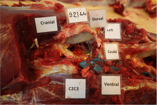
Figure 4.

Presence of blue latex within the vertebral canal at the level of C3. The left cranial and abaxial articular process of the 3rd cervical vertebra was transected with an osteotome to visualize the vertebral canal. In this case, the latex was in contact with the nerve root (*) but a small amount was also present within the vertebral canal (∧), but outside of the dura mater.
