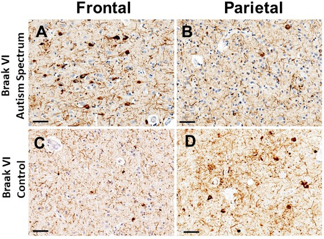
Figure 1.
Figure Legend. Photomicrographs show p-Tau immunoreactive pathologic features in cortical regions of a person who died at age 72 years following new-onset autism spectrum symptoms (A,B), and a control subject, age 76 years at death (C,D). Both individuals had the APOE e4 risk allele, were clinically diagnosed with Probable Alzheimer’s disease, and subsequently had autopsy-confirmed Braak NFT Stage VI pathologic changes. For the individual with autism spectrum symptoms, the tau tangle pathology was more severe in the frontal (Panel A; Brodmann area 9) than parietal (Panel B; Brodmann area 39) cortical region. By contrast, in the individual lacking autism spectrum symptoms, the reverse was true: tau tangle pathology was more modest in the frontal cortex (Panel C) in comparison to that in the parietal cortex (Panel D). Notably, a large proportion of p-Tau-immunoreactive staining in all sections was in neuropil threads rather than intracytoplasmic tangles. Immunohistochemical stains were performed using the PHF-1 antibody (a gift from Dr. Peter Davies), and sections were counterstained with hematoxylin. Scale bars = 50microns.

