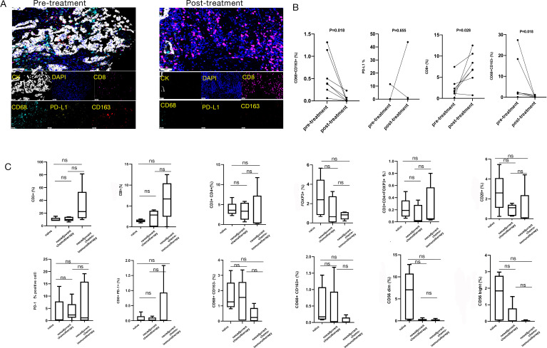
Figure 2.
Multiplex immunohistochemistry assay of immune microenvironment in three different treatment groups. (A) Establishment of a multiplex immunofluorescence staining assay to detect 4 different indices (CD8, CD68, CD163, and PD-L1) of immunomicroenvironment before and after neoadjuvant chemoimmunotherapy. (B) The difference of each index in immune microenvironment before and after neoadjuvant chemoimmunotherapy by Wilcoxon test. CD8+ cytotoxic T cells in the tumor stroma area showed a consistent upward trend after treatment; CD68+CD163- M1 macrophages in the tumor stroma showed a downward trend in each sample after treatment; CD68+CD163+ M2 macrophages in the tumor stroma showed a downward trend in each sample after treatment; PD-L1+ cells in the tumor stroma showed no significant difference before and after treatment (four out of seven patients with PD-L1 TPS < 1% or negative before neoadjuvant chemoimmunotherapy (0, 0.05%, 0.04%, 0), and 5 patients had PD-L1 TPS < 1% or negative after treatment (0, 0, 0, 1.04%, 0)). (C) The differences of immune microenvironment in 3 different treatment groups. There were no obvious differences by multiple comparisons (Mann-Whitney test) among the three groups. ns, P>0.05). (TPS, tumor proportion score)

