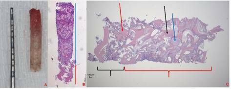
Figure 3. (A) Biopsy sample collected from grafted maxillary sinus. (B) Hematoxylin and Eosin (H&E) stained sections of the biopsy used for histomorphometry analysis. The red line represents the analysis of the linear length of the NB and the blue line represents the measure of the linear length of the GB. (C) The evaluation of the composition of the biopsies at the NB indicated by the black bracket is possible to check only the presence of the bone indicated by the red arrow that was considered the mineralized tissues of these regions. Furthermore, the GB indicated by the red bracket presents the mineralized tissue composed by bone (black arrow) and the DBB remnants (blue arrow).

