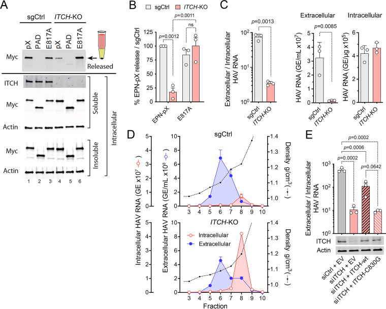
FIG 4.
ITCH depletion impairs the release of EPN-pX and quasi-enveloped eHAV. (A) EPN-pX release from ITCH-KO and sgCtrl cells transfected with EPN-pX. (A, Top) Myc immunoblot of released extracellular EPN-pX protein recovered following centrifugation through a 20% sucrose cushion. (A, Bottom) Immunoblots of soluble intracellular ITCH and soluble and insoluble intracellular EPN-pX. Actin is included in blots as a loading control. (B) Quantitation of EPN-pX and EPN-E817A (Fig. 3) release from ITCH-KO cells relative to release from sgCtrl cells in 3 independent experiments. Percent release was normalized to EPN expression based on the quantitation of soluble intracellular Myc. (C) Intracellular and extracellular HAV RNA abundance in ITCH-KO and sgCtrl cells 24 h postinfection with 18f virus, determined by RT-PCR. Data shown are means ± SD from a representative experiment. The relative abundance of RNA in extracellular fluids (genome equivalents [GE] per mL) versus intracellular viral RNA (GE/μg total RNA) is shown on the right. (D) HAV RNA in fractions of isopycnic iodixanol minigradients loaded with supernatant fluids or lysates of sgCtrl and ITCH-KO cells 5 days postinfection with 18f virus. (E) eHAV release from infected cells depleted of ITCH by transfection of ITCH-specific siRNA and transfected with vectors expressing either wild-type (WT) or catalytically inactive (C830G) ITCH. Also shown are immunoblots of ITCH and actin (loading control). EV, empty vector. Data shown are from 3 technical replicates in a representative experiment. Statistical comparisons were done by two-way ANOVA.

