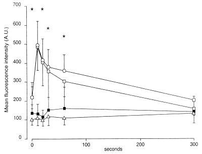
FIG. 5.
Time course of f-MLP-induced changes in cellular F-actin contents in human granulocytes. After preincubation of granulocytes with preopsonized Y. enterocolitica and removal of nonadherent bacteria, the pellet was stimulated with 100 nM f-MLP. After permeabilization, granulocytes were stained with fluorescein phalloidin. Fluorescence intensity was measured by flow cytometry. Results are expressed as means (± standard errors) of values determined for fluorescence intensity. ∗, values for pYV+ Y. enterocolitica and Y. enterocolitica W22703(pGC1152) YadA+ YopH− that were significantly different from values for pYV− Y. enterocolitica (P < 0.05, paired Student’s t test). Normal controls (□) and granulocytes preincubated with pYV− Y. enterocolitica (○), pYV+ Y. enterocolitica (■), or Y. enterocolitica W22703(pGC1152) YadA+ YopH− (▵) were tested. A.U., arbitrary units.

