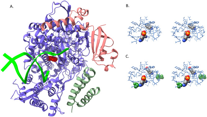
Figure 8.
(A) RNA-dependent RNA polymerase (blue) complex formed with non-structural protein 7 (green) and non-structural protein 8 (salmon). Primer and template RNA strands (lime green) are depicted as ribbons extending from the active site. The mutated residue (F694Y) is rendered as a red CPK model. (B) Stereo pair view of the active site with bound remdesivir molecule. Both remdesivir and F694 are rendered as CPK models, and their atoms are color-coded by atom type as described in Fig. 5. (C) Stereo pair view of active site with bound remdesivir in the F694Y mutant. Residues F480 and V557, which impart inhibitor resistance in SARS-CoV-1 polymerase, are rendered as green CPK models to illustrate their locations relative to remdesivir. F480 is on the right side of the image and V557 is in the lower left region. The orientation and color-coding are identical to that in Fig. 8B. The mutant homology model has not been subjected to further structural refinement and this image clearly displays steric clashes generated by substitution of the tyrosine side chain. Models in panels A, B and C are all based on the cryo-electron microscopy structure PDB ID: 7BV2.

