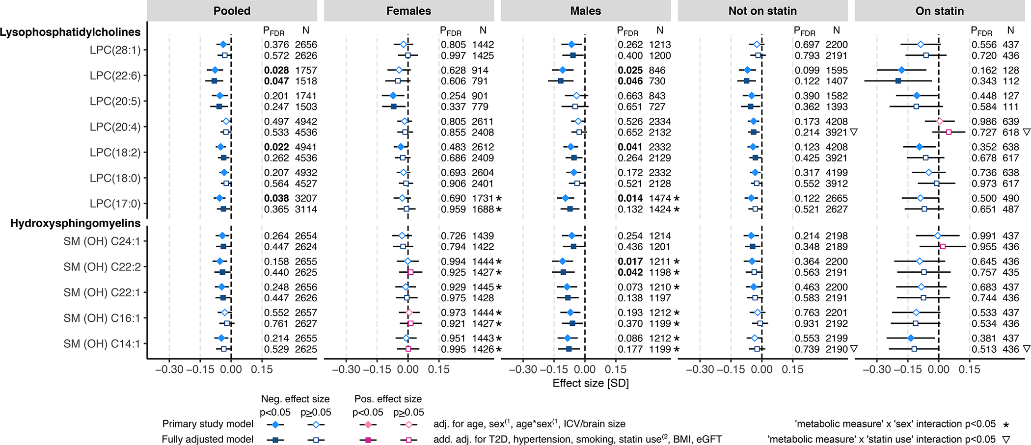
Figure 3. WMH associations with circulating levels of lysophosphatidylcholines (LPC) and hydroxylated sphingomyelins (SM (OH)).

The associations between WMH and circulating metabolomic measures were determined using linear regression. The primary study models (diamonds) were adjusted for age and intracranial volume or brain size, and, in the pooled sample and in statin treatment-stratified subsamples also for sex and age-by-sex interaction(1. The fully adjusted models (squares) were additionally adjusted for type 2 diabetes (T2D), hypertension, current smoking status, body mass index (BMI), and estimated glomerular filtration rate (eGFR), and, in the pooled sample and in sex-stratified subsamples, also for statin treatment(2. Where relevant, all models were adjusted for fasting duration, time between blood sampling and brain MRI, and possible cohort study-specific covariates. Blue color indicates negative effect size and red color indicates positive effect size. Error bars indicate 95% confidence intervals. P-values below the threshold for multiple testing correction (pFDR<0.05) are indicated with bold font. FDR indicates false discovery rate; LPC, lysophosphatidylcholine; and SM (OH), hydroxylated sphingomyelin
