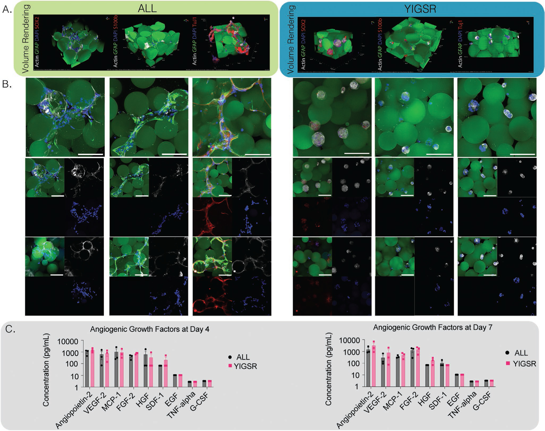
Figure 5.

A) NPCs were cultured in vivo for 4 days. Confocal images were taken at 40× and volumed renderings or max intensity projections (MIPs) were displayed for ALL and YIGSR conditions. Scaffolds were stained for SOX2 for stemness, Tuj1 for neurogenesis, or S100b for astrogliosis, scale bar is 100 μm. B) Representative images of ALL and YIGSR conditions stained for differentiation markers, scale bar is 100 μm. C) Luminex multiplex immunoassay for angiogenic markers at day 4 and day 7 for ALL or YIGSR conditions, no statistical difference was observed between days.
