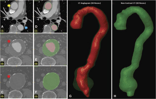
Figure 1.

A-F. Axial Slices obtained from a CTA and noncontrast CTscan with overlaying manually segmented labels. The lumen is illustrated in black and is typically surrounded by the outer wall in grey. In the abdominal region, the grey label includes the intra-luminal thrombus, if present. G–H. 3D-reconstructed volumes representing the aortic lumen (black) and wall structure (grey), which contains the intra-luminal thrombus, are generated from the masks.
