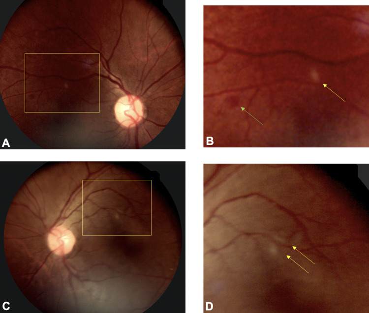
Fig 2. Fundus photographs of a COVID-19 patient with subtle bilateral retinopathy.
(A) Fundus photo of the right eye (A) shows cotton wool spots and retinal hemorrhage (yellow inset). (B) A higher magnification view of the inset shows peri-arterial cotton wool spot (yellow arrow) and dot blot hemorrhage green. (C). Fundus photo of the left eye shows focal peri-arterial cotton wool spots (inset). (D) A higher magnification view of the cropped inset shows two cotton wool spots adjacent to retinal arteriole.

