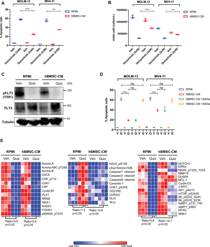
Figure 1. Conditioned media of human bone marrow stromal cells (hBMSCs) prevents apoptosis in FLT3-ITD acute myeloid leukemia (AML) cells upon FLT3 inhibition.
(A, B), FLT3-ITD AML cell lines, MOLM-13 and MV4-11, were treated with either DMSO (veh) or quizartinib with indicated doses for 48 hr in regular media (RPMI) or 50% conditioned media of human BM stromal cells in RPMI (hBMSC-CM). (A) Apoptotic cells were detected by 7-AAD/Annexin V staining post-treatment. (B) Cell viability was measured based on propidium iodide (PI) exclusion after 5 days of rebound growth post-treatment (n = 3). (C), MOLM-13 cells were harvested after 15 hr of indicated treatments (Quiz: 3 nM quizartinib) to determine FLT3 activation by Western blotting. (D), MOLM-13 and MV4-11 cells were treated with either vehicle (V) or quizartinib (Q) for 48 hr in RPMI, hBMSC-CM, RPMI supplemented with heavy fraction (>30 kDa) of hBMSC-CM or light fraction (<30 kDa) of hBMSC-CM, followed by detection of apoptotic cells. (E), MOLM-13 cells were harvested after 16 hr of treatments as indicated and subjected to reverse-phase protein array analysis to explore the expression/activation levels of a wide range of proteins.
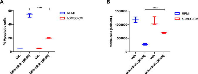
Figure 1—figure supplement 1. Human bone marrow stromal cells (hBMSC-CM) provide protection against apoptosis in FLT3-ITD acute myeloid leukemia (AML) cells following FLT3 inhibition by gilteritinib.
Figure 1—figure supplement 2. Human bone marrow stromal cell (hBMSC-CM)-mediated protection does not coincide with alterations of the cell cycle.
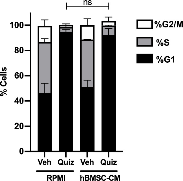
Figure 1—figure supplement 3. Human bone marrow stromal cell (hBMSC-CM) from HS-27A does not provide protection against apoptosis in FLT3-ITD acute myeloid leukemia (AML) cells following FLT3 inhibition.
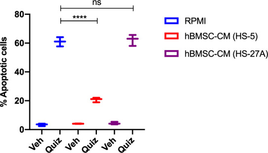
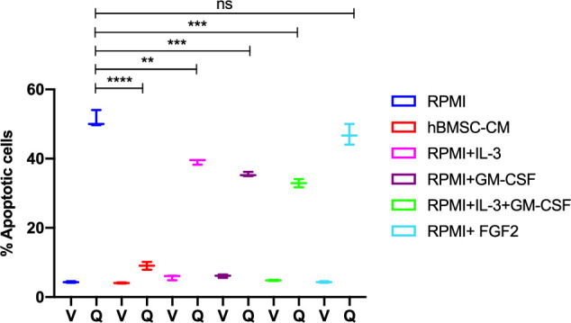
Figure 1—figure supplement 4. Human bone marrow stromal cell (hBMSC-CM)-mediated protection is partially mimicked by BM-enriched cytokines.
