
Figure 2.
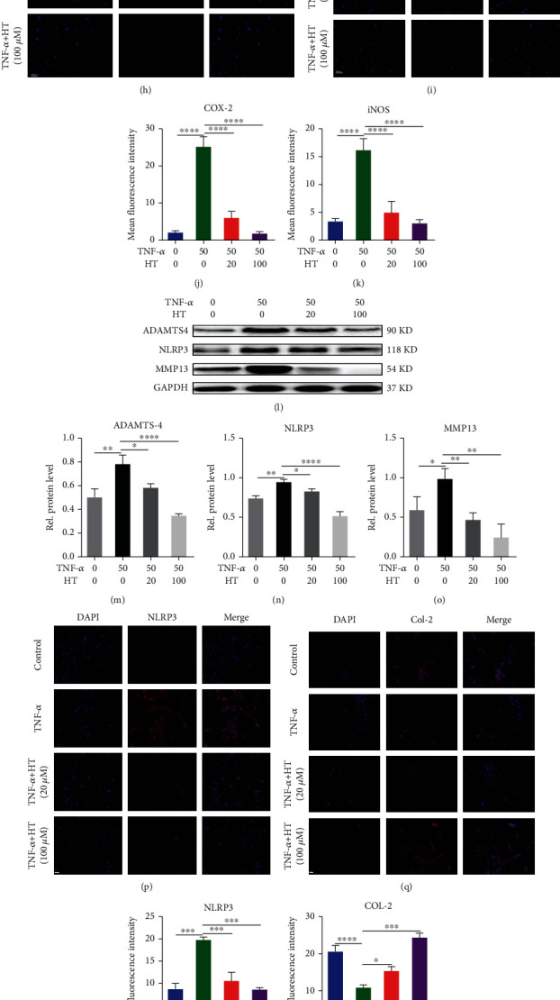
HT protects the main components of the IVD by alleviating inflammation and mitigating ECM degradation. Note: HNPCs were incubated for 24/48 h with 50 ng/mL TNF-α and 20 or 100 μM HT or were left untreated. The expression of iNOS (a), COX-2 (b), MMP-13 (c), and ADAMTS-4 (d) was assayed by qRT–PCR. Protein levels of iNOS (e, f) and COX-2 (e, g) were assessed by WB. Immunofluorescence staining was used to assess the COX-2 (h, j), and iNOS (i, k) levels in HNPCs treated with TNF-α and HT. Scale bar: 50 μm. Western blot detection for the expression of ADAMTS-4 (l, m), NLRP3 (l, n), and MMP13 (l, o) in each group. Immunofluorescence staining of NLRP3 (p, r), col-2 (q, s), aggrecan (t, w), and MMP-13 (u, v) in HNPCs. Scale bar: 50 μm.