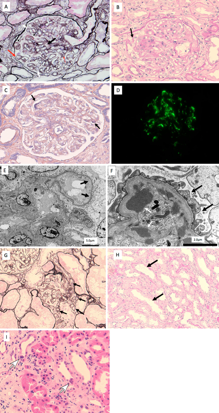
Figure 2.

Kidney biopsy findings. A (periodic acid methenamine silver stain): The kidney biopsy showed features consistent with thrombotic microangiopathy, with endothelial swelling, a subendothelial insudative lesion (thin black arrows), duplication of the glomerular basement membrane (white arrow), mesangiolysis (thick black arrow), endothelial swelling (thick red arrow), and vacuolar degeneration of the podocytes (thin red arrows), although overt thrombosis was not noted in the small arteries or glomeruli. B (Periodic acid Schiff stain): Massive hyaline degeneration of the podocytes (black arrow) was also evident, suggesting podocytopathy. C: The subendothelial insudative lesions (black arrows) were considered fibrin material that was positive for phosphotungstic acid hematoxylin (PTAH) staining. D: In the mesangial areas, immunofluorescence staining was positive for IgM. E: Electron microscopy showed marked widening of the subendothelial spaces (black arrows). F: Electron microscopy showed massive electron-dense material in the subendothelial space (white arrow). Podocyte foot process effacement (black arrows) was focal and mild. G (Periodic acid methenamine silver stain): Polar vasculosis was observed around the glomerular vascular pole (black arrows). H (Periodic acid Schiff stain): Brush border loss of proximal tubular epithelial cells (black arrows). I (Hematoxylin and Eosin staining): Multinuclear formation of distal tubular epithelial cells (white arrows) was noted.
