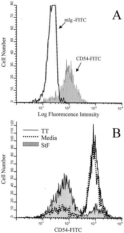
FIG. 11.
Effects of STF on Mφ expression of CD54. PBMC from a vaccinated volunteer were stained immediately after isolation (A) or cultured overnight in the absence (Media) or presence of TT (2 μg/ml) or STF (2 μg/ml) (B). The cells were labeled with control antibodies (FITC-labeled mouse IgG [mIg-FITC]) or CD54-FITC, as described in Materials and Methods. The histograms shown correspond to cells gated on the Mφ region, characterized by high forward light scatter versus high side light scatter. (A) Dotted area represents cells expressing CD54 immediately after isolation. (B) Shaded area represents cells expressing CD54 after incubation with STF. Shown are results from an individual representative of five volunteers (four vaccinated, one unvaccinated) evaluated in two independent experiments with similar results.

