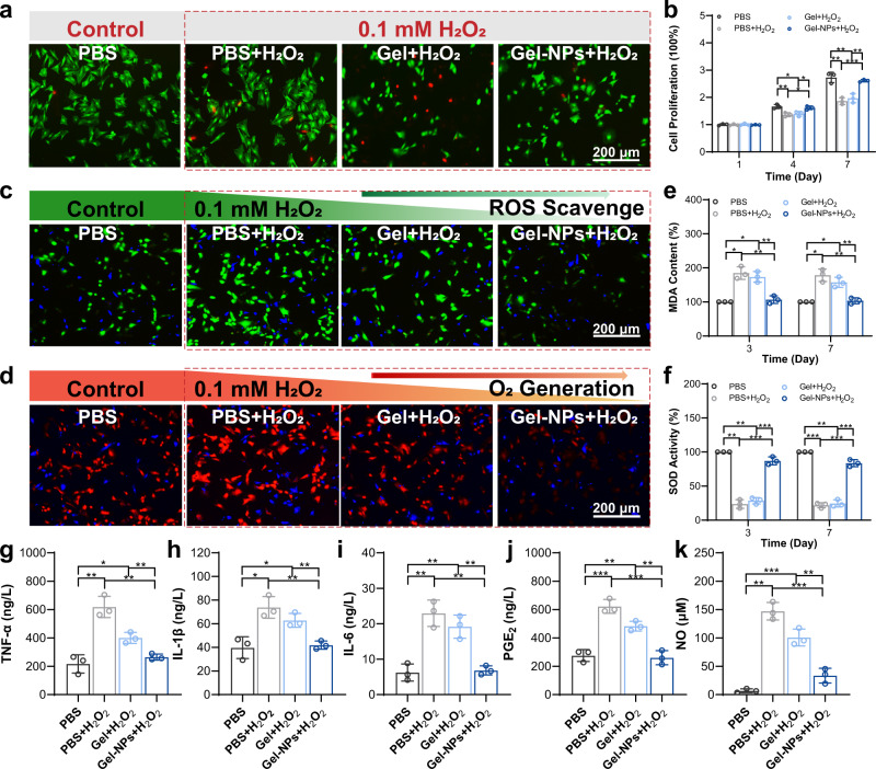
Fig. 4. In vitro studies of BMSCs treated with the hydrogel.
a Calcein-AM/propidium iodide (PI) staining of BMSCs after treatment with PBS, PBS + H2O2, Gel+H2O2, and Gel-NPs+H2O2. b Cell proliferation of BMSCs in different groups at the 1st, 4th, and 7th days. c ROS scavenge ability validated by a ROS probe (DCFH-DA) after different treatments. Green fluorescence from DCFH-DA indicates the presence of ROS. d Intracellular O2 generation assay monitored by an O2 probe [Ru(dpp)3Cl2]. Red fluorescence from Ru(dpp)3Cl2 is quenched by O2. e MDA activity of BMSCs after different treatments. f SOD activity of BMSCs after different treatments. g–k Expression of inflammatory mediators of BMSCs after different treatments including TNF-α (g), IL-1β (h), IL-6 (i), PGE2 (j), and NO (k). These data are presented as mean values ± SD (n = 3 independent experiments). Statistical significance was determined by two-tailed t test. *P < 0.05, **P < 0.01, and ***P < 0.001. Source data and exact P values are provided as a Source Data file.

