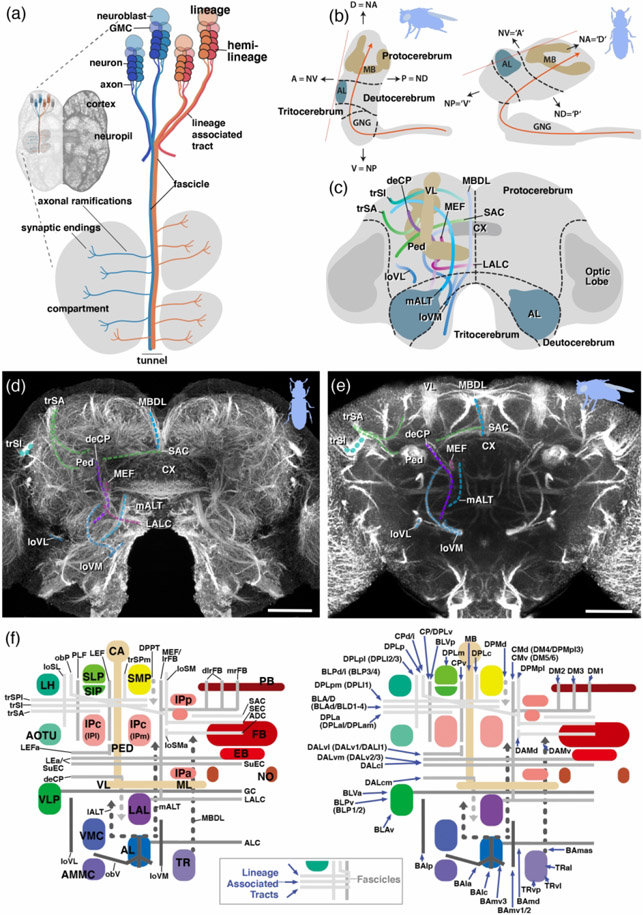
Figure 1: Fundamentals of insect brain anatomy.
(a) Schematic of larval brain illustrating the features of neural lineage formation [lineage, hemilineage, neuroblast, ganglion mother cell (GMC), neuron, lineage associated tract] and the spatial relationship between lineage associated tracts and cortex, neuropil and neuropil compartments; (b) Schematics of brains of D. melanogaster and T. castaneum (lateral view), showing neuraxis (orange line), arrangement of brain neuromeres [protocerebrum, deutocerebrum, tritocerebrum, gnathal ganglia (GNG)] and angle of frontal plane (orange dashed line) relative to neuraxis. (c) Schematic of insect brain (anterior view, shape of adult T. castaneum brain) with neuromeres and assemblage of representative neuropil fascicles in different colors. (d, e) Z-projections of frontal confocal section of adult T. castaneum (d) and D. melanogaster brain (e) labeled with global neuronal marker (T. castaneum: Tubulin; D. melanogaster: Neuroglian). Segments of homologous fascicles shown in the schematic (c) are shaded and annotated. (f) Schematic T. castaneum brain representations of fascicles and compartments (left) as well as tracts (right) now described and implemented in this study. Colours as in the models. For abbreviations see Table 2. Scale bars: 25μm (d, e). Animal icons were taken from phylopic.org

