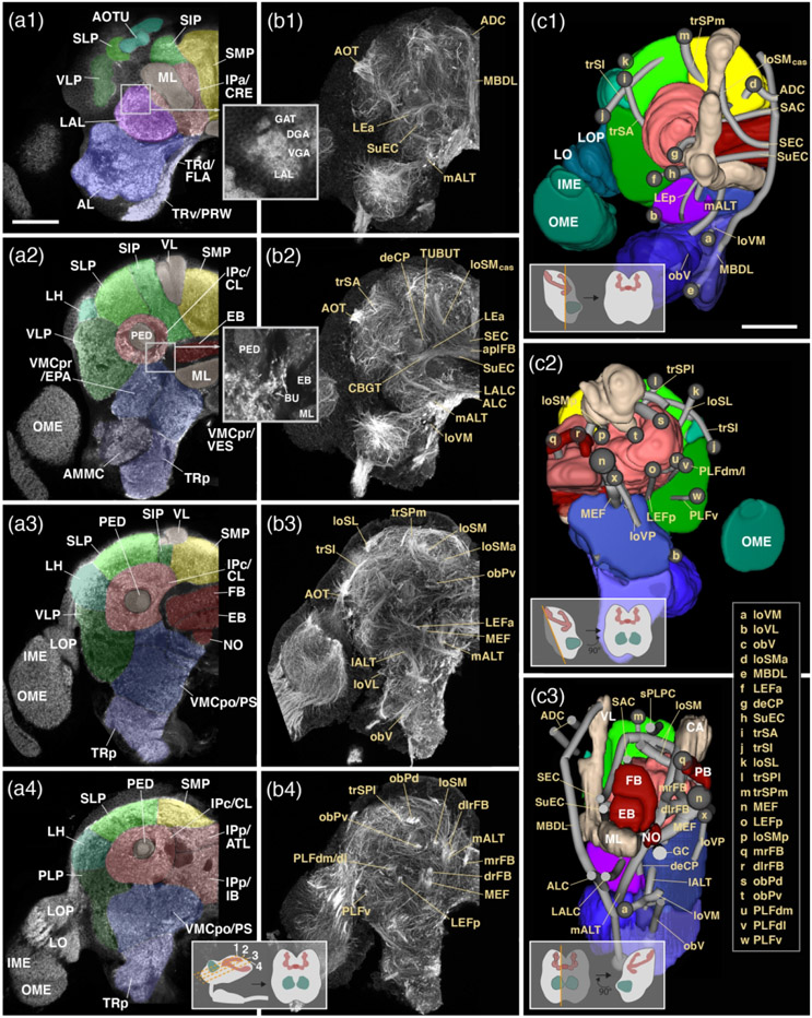
Figure 3: Structure of the adult brain of T. castaneum: Neuropil compartments and fascicles.
(a1-4): Z-projections of frontal confocal sections of adult brain hemisphere labeled with an antibody against synapsin. The four z-projections represent brain slices of approximately 8–10mm thickness and are arranged in anterior (a1) to posterior (a4) sequence at levels shown in inset at bottom of a4. Compartments are annotated with white letters. (b1-4): Z-projections of frontal sections of larval brain hemisphere labeled with Tubulin antibody, arranged as explained for the a1-4 series. Fascicles are annotated in beige lettering. (c1-3): Digital 3D models of one brain hemisphere in anterior view (c1), posterior view (c2) and lateral view (c3). Insets show not easily visible details about the lateral complex and its neuropils. As explained for Figure 2d1-5, compartments are color coded, but parts of compartments facing the viewer are cut away to allow for observing fascicles. Starting points of fascicles are shown has single letter-containing spheres; fascicles themselves are annotated in beige lettering. For all abbreviations see Table 2. Scale bars: 50μm (a1-4, b1-4; c1-3).

