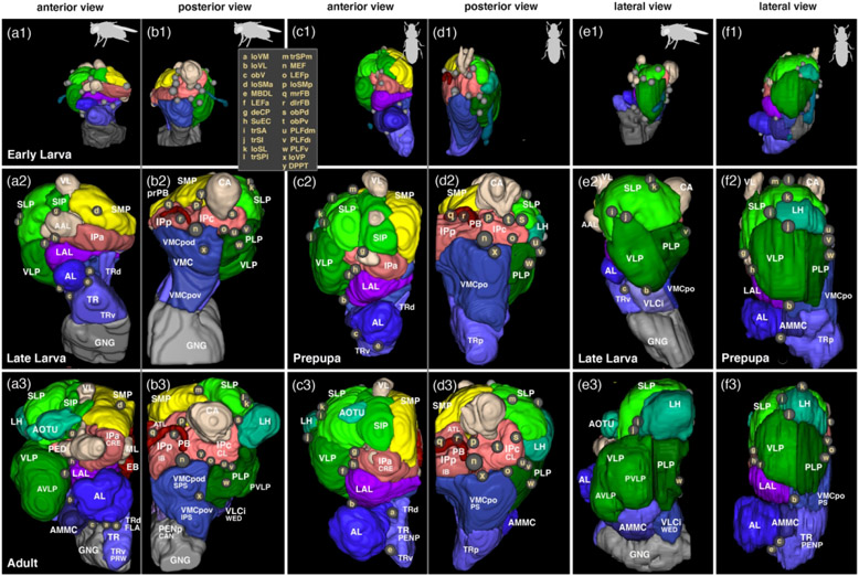
Figure 7: Comparative growth of brain compartments in D. melanogaster and T. castaneum.
Panels show digital 3D models of one brain hemisphere of early larva (top row), late larva/prepupa (middle row) and adult (bottom row) in anterior (a1-3, c1-3), posterior (b1-3, d1-3) and lateral view (e1-3, f1-3), all to scale. Neuropil compartments are rendered in the same colors as in previous figures (Figures 2-5) and are annotated in white lettering. As in previous figures (Figures 2-4), small spheres annotated in beige single letters represent locations where neuropil fascicles enter the neuropil. For correspondence of single letters with fascicle names see inset in b1 and c1. For all abbreviations see Table 2. The D. melanogaster larval models are based on Cardona et al. (2010) and the adult model is based on Pereanu et al. (2010). Animal icons were taken from phylopic.org

