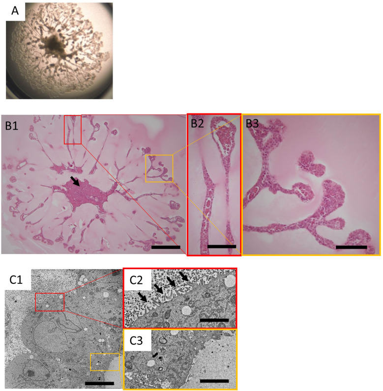
Fig. 1.
Histology of the organoids derived from the KS cells.
A: Stereomicroscopic image of organoids derived from KS cells on day 20 in the 24-well transwell insert. B1: Histology of organoids from KS cells on day 20. The cells were mostly necrotic at the center of the culture (arrow). Haematoxylin and eosin (HE) stained; bar=600 µm. B2, B3: Magnified images of B1. The tubular structures of the organoids were generally lined with a single layer of cells. The tip of the tubular structure was bulged, and small cell populations were often trapped within the tubular structure. HE stained sections; bar=150 µm. C1: Electron microscopic image of organoids derived from KS cells on day 20; bar=5.0 µm. Magnified images of the luminal side (C2) and outer side (C3) of the tubular structure; bar=1.25 µm.

