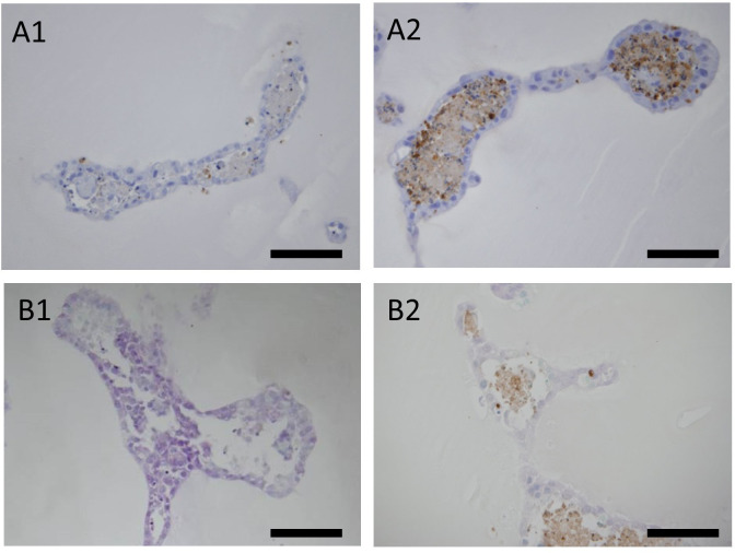
Fig. 5.
Cisplatin-induced apoptosis in organoids derived from KS cells.
A: Immunohistochemistry of cleaved caspase 3 (apoptosis marker); bar=100 µm. A1: Tubular structures of organoids not exposed to cisplatin. A2: Tubular structures of organoids exposed to 20 µM cisplatin for 144 h. Positive sites were often observed inside the tubular structure. B: TUNEL assay of organoids derived from KS cells (bar=100 µm). B1: Tubular structures of organoids not exposed to cisplatin. B2: Tubular structures of the organoids exposed to 10 µM cisplatin for 144 h. Positive sites were observed mainly inside the tubular structures.

