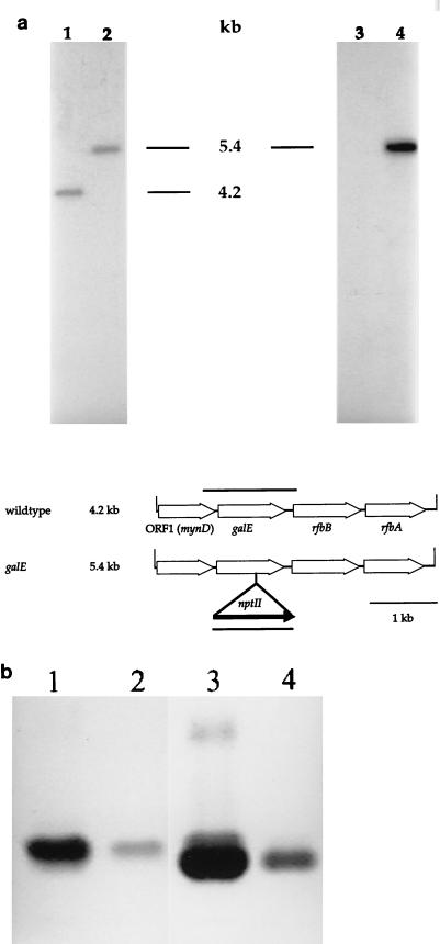
FIG. 2.
Analysis of N. meningitidis MA-1 galE. (a) Southern blot analysis of genomic DNA from N. meningitidis MA-1 galE. Genomic DNAs isolated from N. meningitidis MA-1 and MA-1 galE were digested with DraI and transferred to a nylon membrane after electrophoresis. Duplicate filters were probed with either a galE gene probe (lanes 1 and 2) or a kanamycin cassette probe (lanes 3 and 4). The hybridization patterns indicated the incorporation of the kanamycin cassette in the galE open reading frame. A genetic map of the N. meningitidis MA-1 galE mutant is shown below. The MA-1 galE gene was cloned on a 4.2-kb DraI fragment. This fragment had four open reading frames, as designated. To construct MA-1 galE, a kanamycin cassette from pBSL14 was inserted into the unique MunI site 647 bases into the galE coding region. This construct was introduced into the chromosome of MA-1 by allelic replacement. Open arrows, meningococcal open reading frames; solid arrow, kanamycin resistance cassette. (b) SDS-PAGE analysis of MA-1 and MA-1 galE LOS. The wild-type LOS migrated as a single band. The LOS from the galE mutant gave two distinct bands, of which the upper band comigrated with the wild-type LOS molecule. The lower band is the Hex-Hep2-GlcNAc-Kdo2-lipid A structure. Lanes 1 and 2, MA-1 LOS (2.0 and 0.2 μg, respectively); lanes 3 and 4, MA-1 galE LOS (2.0 and 0.5 μg, respectively).

